
The Human BioMolecular Atlas Program (HuBMAP)
The Human BioMolecular Atlas Program (HuBMAP)
Image of the Week
Some of the most amazing things to come out of the HuBMAP Consortium are the images of healthy human tissues generated by our researchers.
Here, we collected them in one place to celebrate the work of these talented individuals.
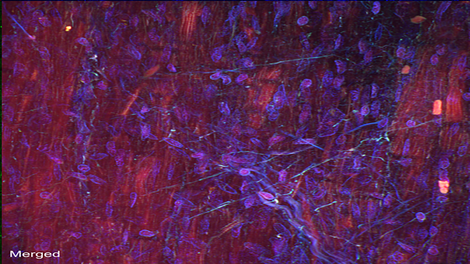
Autofluorescence image capturing heart cells (red), nuclei (blue), and dense fibers of the heart (green), courtesy of Dr. Seth Currlin at University of Florida
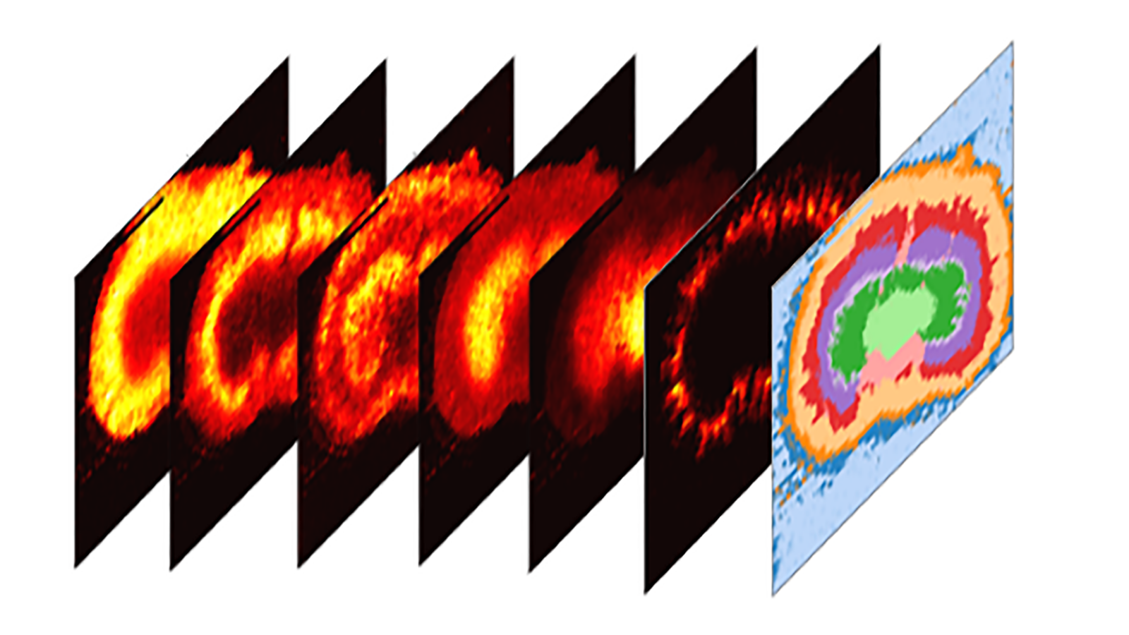
Maps of imaging mass spectrometry data from rat brain, courtesy of Hang Hu at PNNL
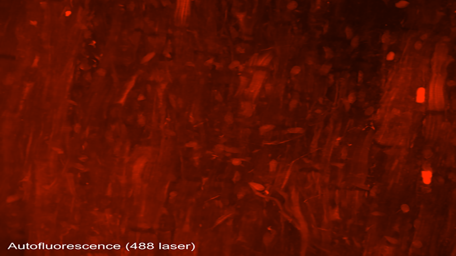
An autofluorescence image of cells that make up the heart/cardiac muscle, from Dr. Seth Currlin of University of Florida
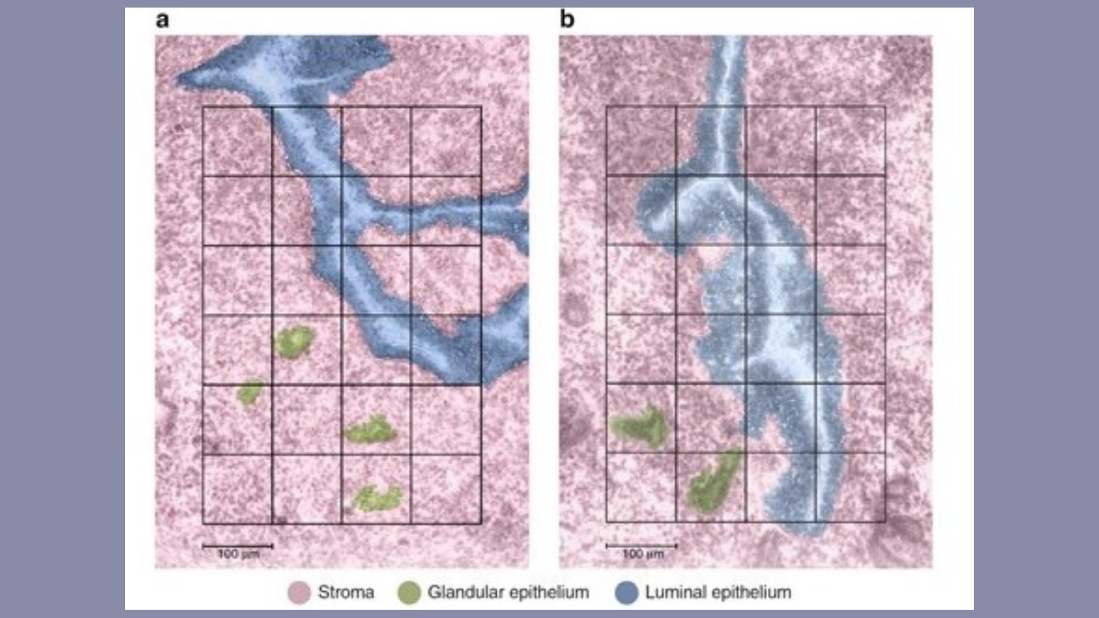
The lining of the uterus & fetal cells within & around maternal spiral arteries, courtesy of Dr. Michael Angelo at Stanford
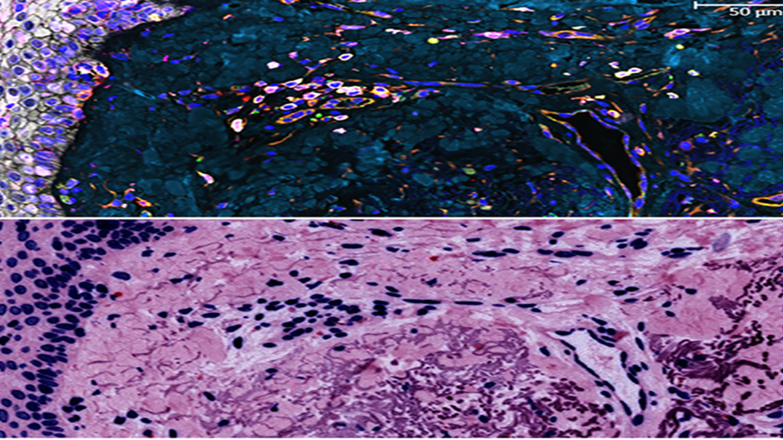
Proteins in the epidermis, the outermost skin layer, courtesy of Dr. Fiona Ginty at GE Research
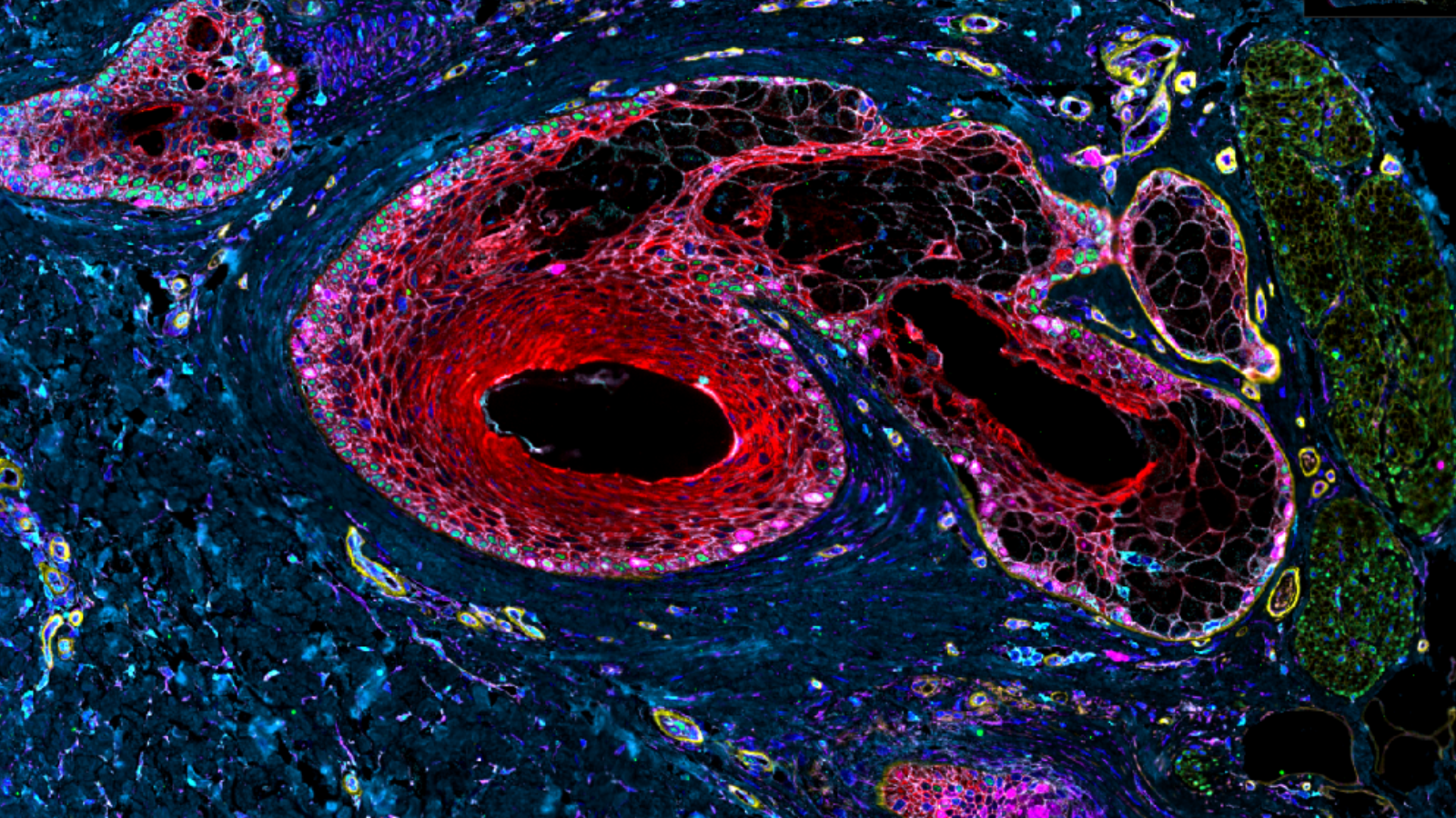
Image shows 9 markers of a follicle in skin, courtesy of Dr. Fiona Ginty at GE Research
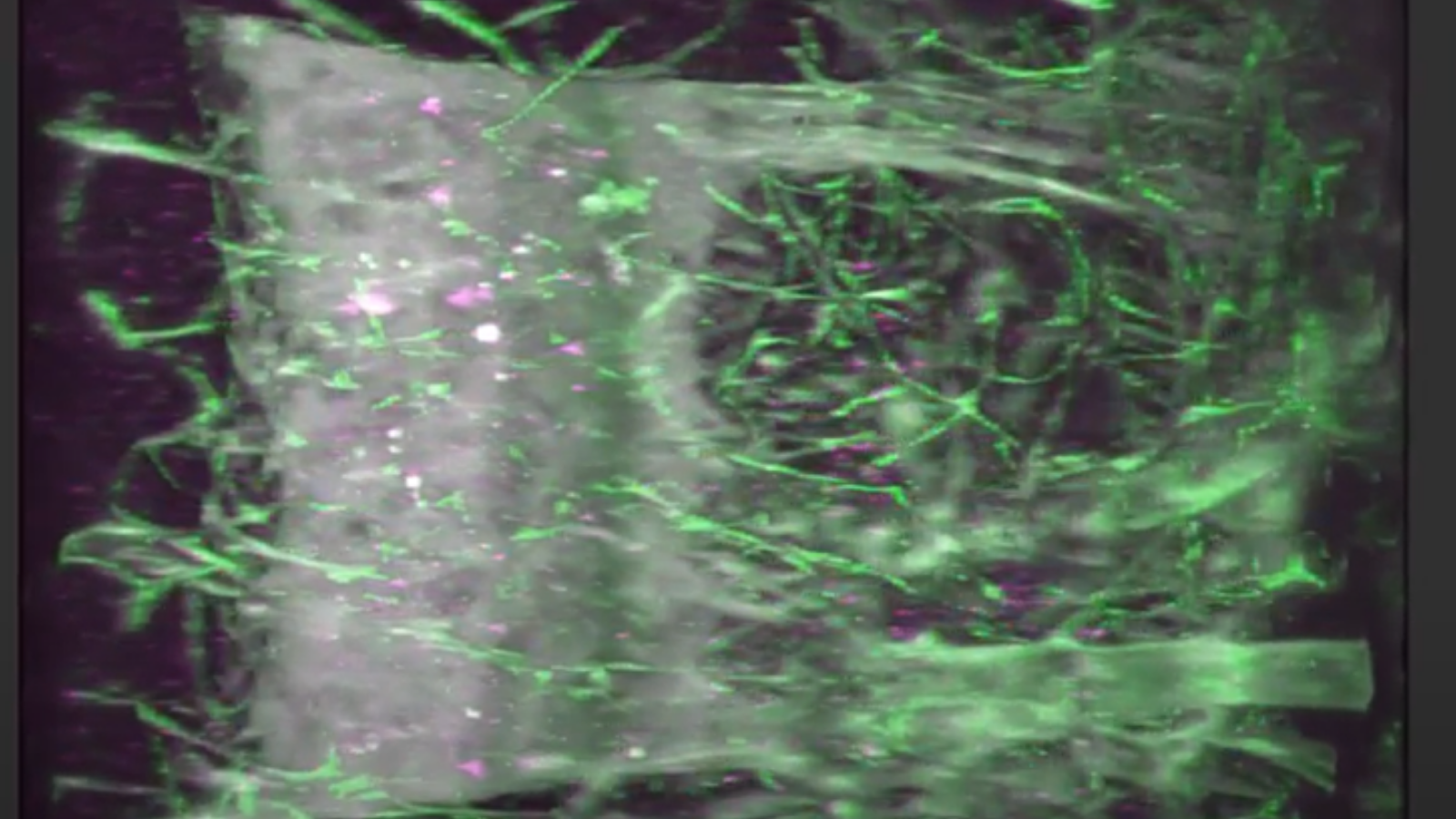
lightsheet image from University of Florida's, Seth Currlin, showing the neural network (green) within a human thymus, where the cells that fight infection mature.
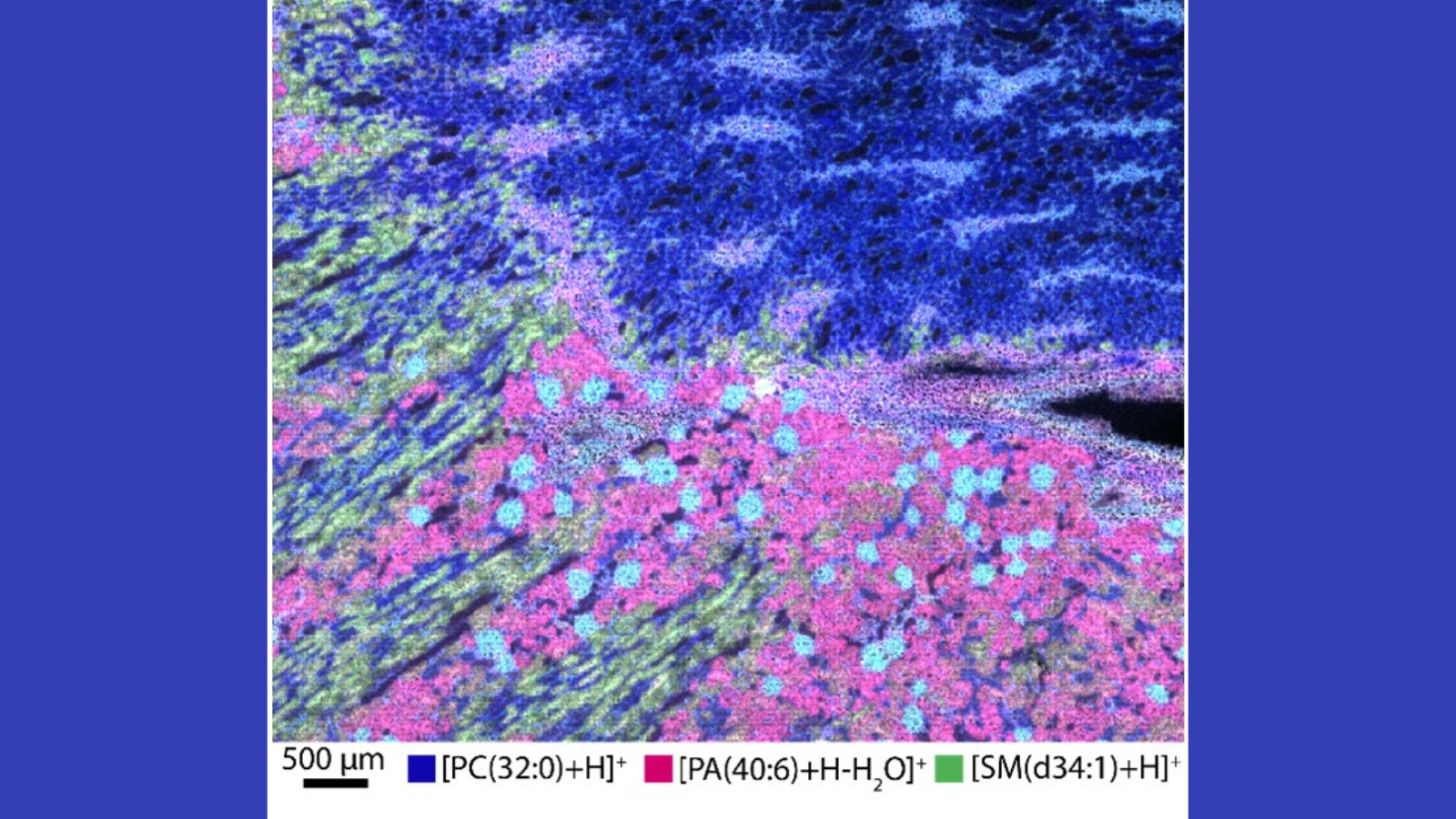
MALDI mass spectrometry image that shows where three kinds of lipids are in different parts of a human male kidney, courtesy of Dr. Elizabeth Neuman of Vanderbilt
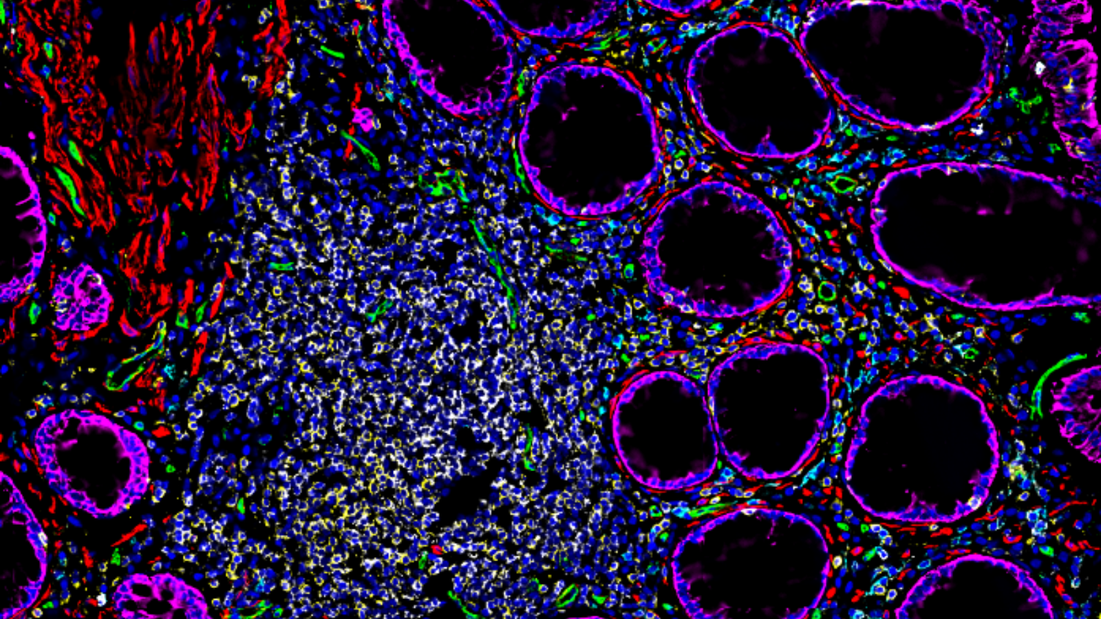
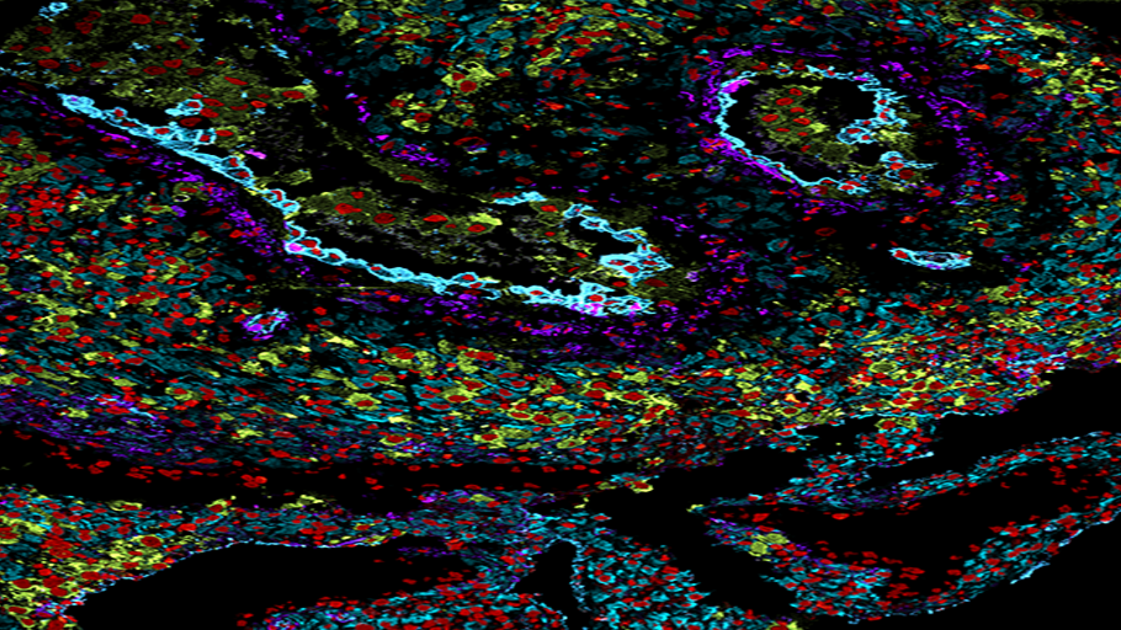
7 proteins in a section of healthy human colon tissue, courtesy of Dr. John Hickey at Stanford
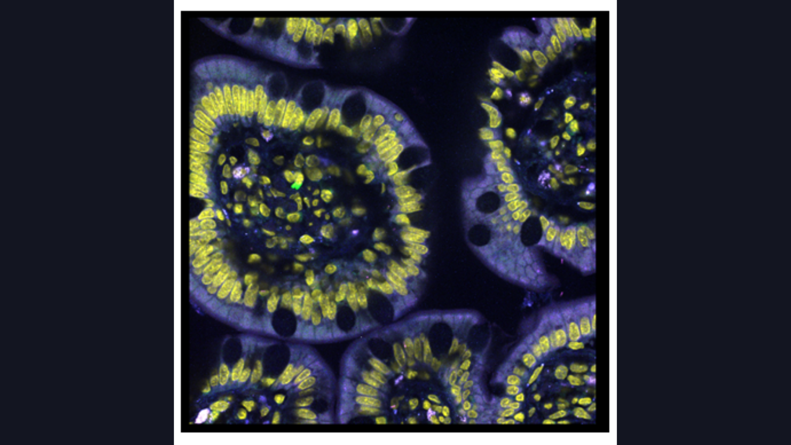
RNA transcripts in sections of the small intestine, courtesy of Dr. Long Cai at Cal Tech
automated mass spectrometry imaging of the proteins in the mouse uterus, courtesy of Dr. Kristin Burnum-Johnson at PNNL
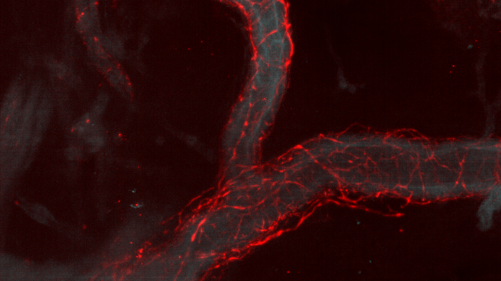
A human splenic blood vessel (cyan) surrounded by the tyrosine hydroxylase matrix (red) from axons near the blood vessel, courtesy of Dr. Seth Currlin, University of Florida
