

The Human BioMolecular Atlas Program (HuBMAP)
The Human BioMolecular Atlas Program (HuBMAP)
Image of the Week
Some of the most amazing things to come out of the HuBMAP Consortium are the images of healthy human tissues generated by our researchers.
Here, we collected them in one place to celebrate the work of these talented individuals.

Cell DIVE image of DNA damage in skin cells from sun exposure courtesy of Dr. Liz McDonough at in the Ginty lab at GE Research

CODEX image of the human retina courtesy of Dr. Angela Kruse at Vanderbilt University

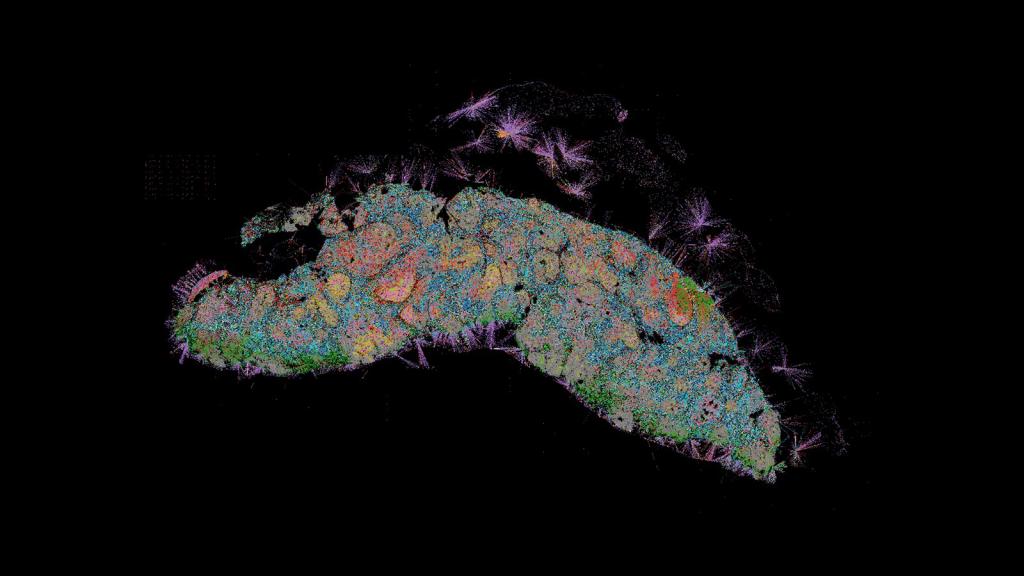
CODEX image of two lymph nodes, courtesy of Archie Enninful in Dr. Rong Fan's lab at Yale University

CellDIVE image of lung courtesy of Dr. Gloria Pryhuber at URMC

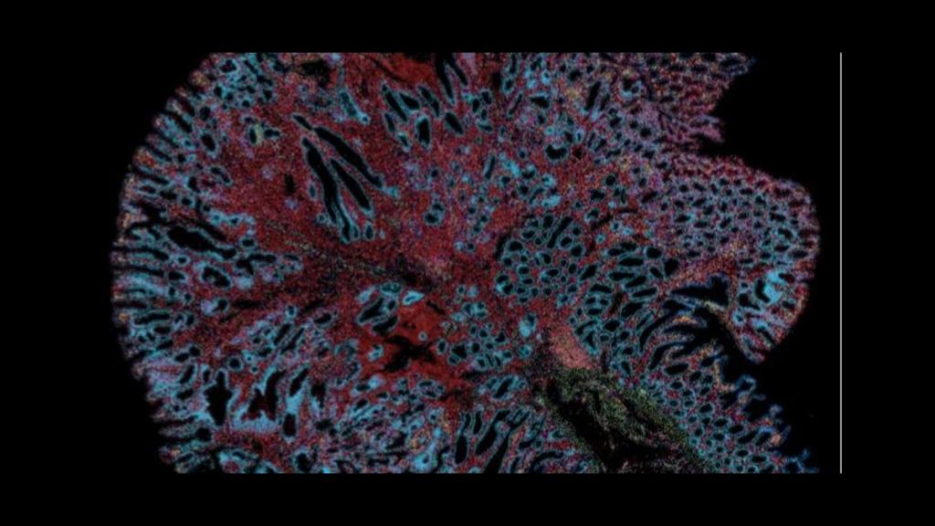
CODEX image of intestine courtesy of Dr. John Hickey from Stanford and Duke Universities

CellDIVE image of glands in skin, courtesy of Dr. Liz McDonough from GE

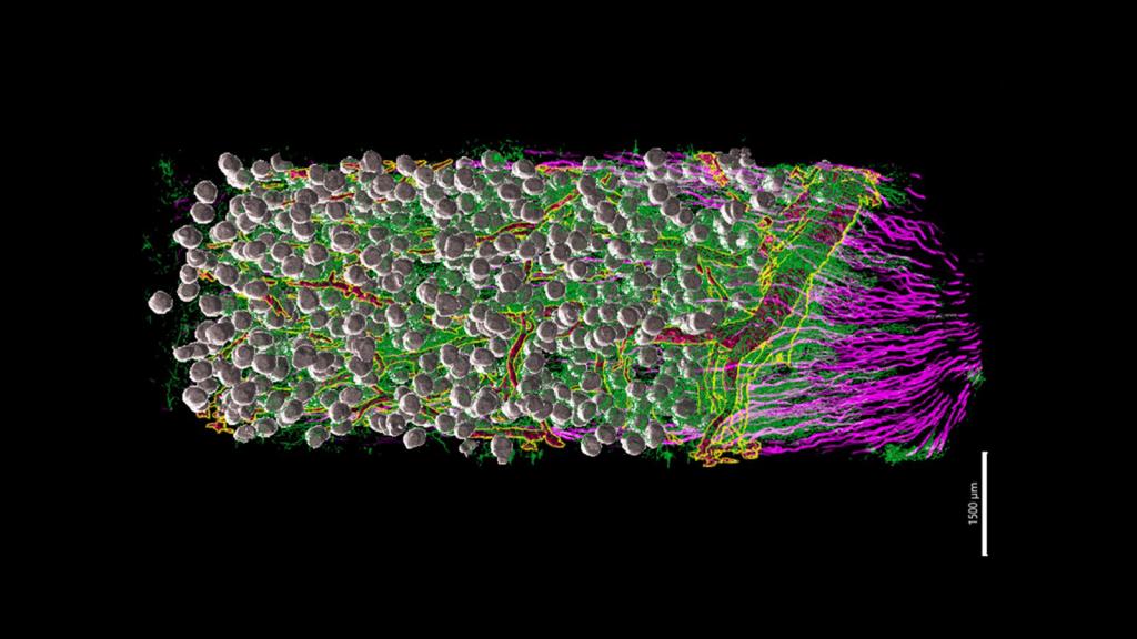
Left panel - CODEX of bone marrow, right panel - visualization of neighborhoods within the bone marrow sample, courtesy of Kai Tan's lab at UPENN
CODEX image of a lymph node, courtesy of Archie Enninful of Yale
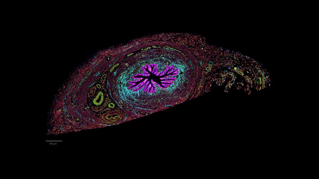
CODEX image of the isthmus, the connection between the uterus and Fallopian tube, courtesy of Dr. Kate O'Neill at UPENN
Xenium image of intestines courtesy of Dr. Chenchen Zhu of Mike Snyder's lab at Stanford University
Lightsheet Microscopy image of kidney courtesy of Liam McLaughlin in Dr. Sanjay Jain's lab at WUSTL
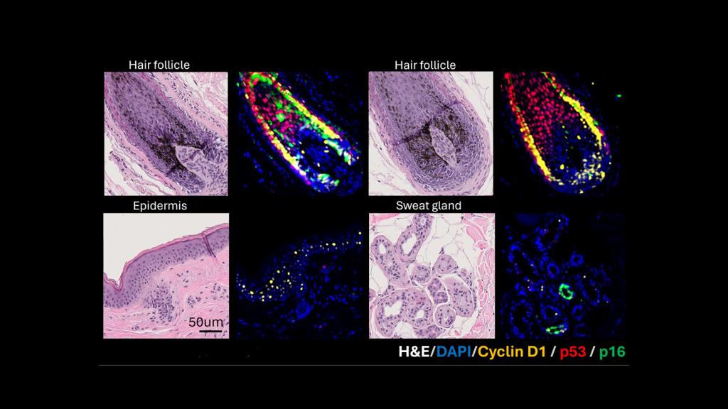
H&E and MxIF images of hair follicles, sweat gland, and epidermis in human skin, courtesy of Dr. Pei-Hsun Wu at JHU.
