
The Human BioMolecular Atlas Program (HuBMAP)
The Human BioMolecular Atlas Program (HuBMAP)
Image of the Week
Some of the most amazing things to come out of the HuBMAP Consortium are the images of healthy human tissues generated by our researchers.
Here, we collected them in one place to celebrate the work of these talented individuals.
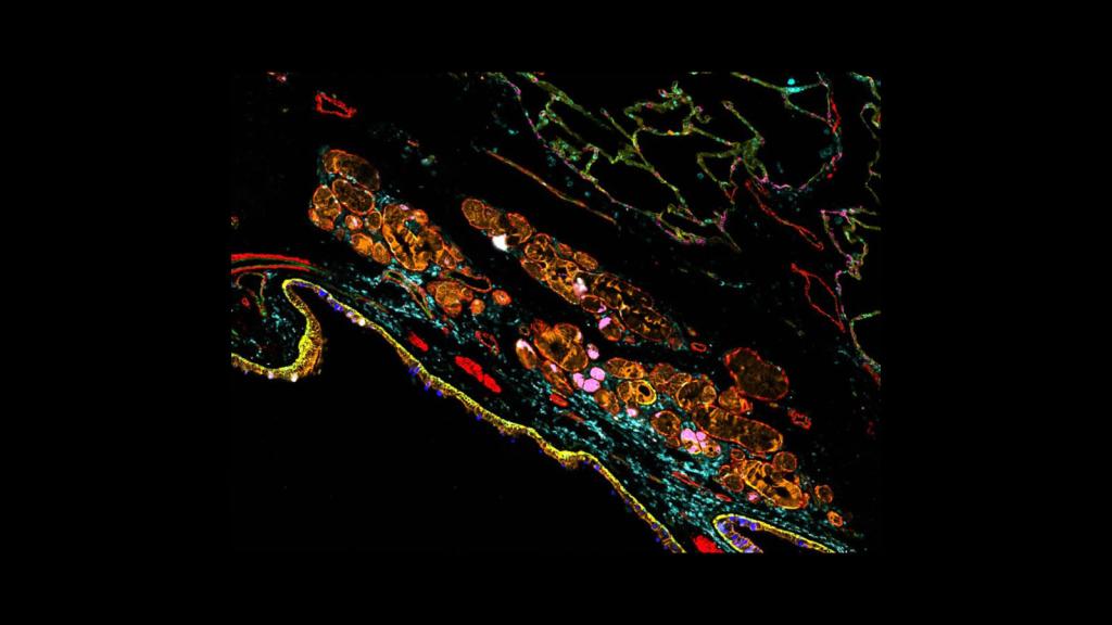
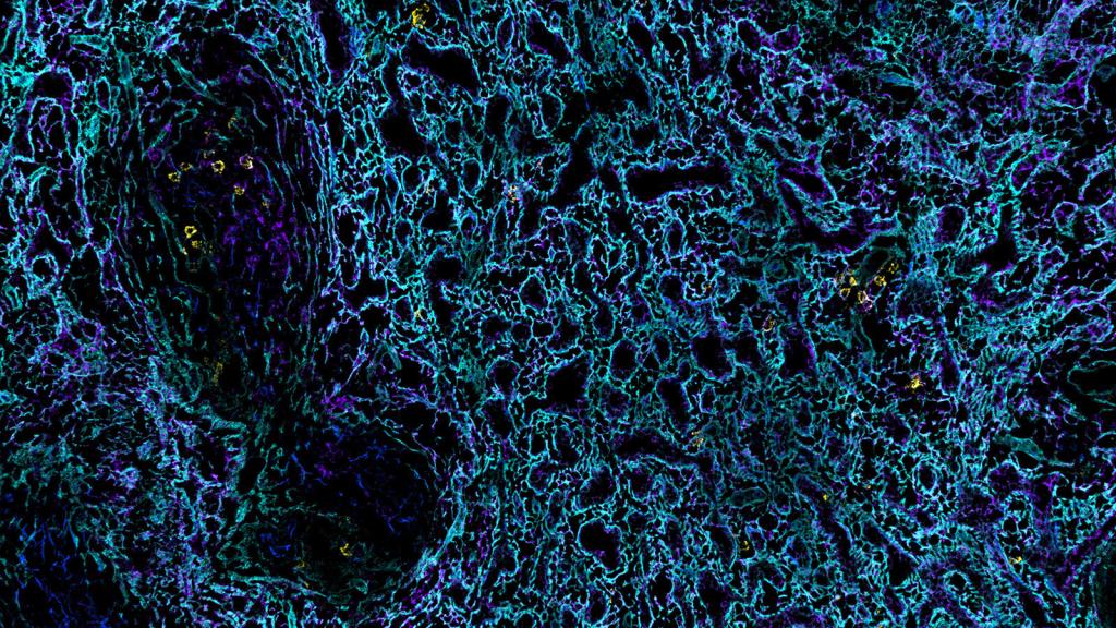
Phenocycler Fusion image by Drs. Gloria Pryhuber and Jeffery Purkerson, at the University of Rochester Medical Center, of the surface of the lung
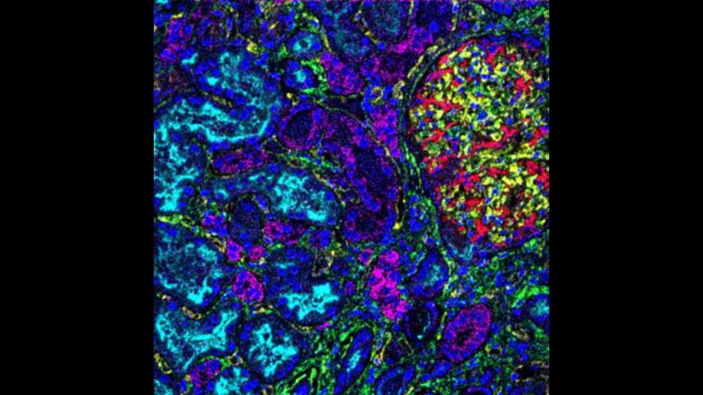
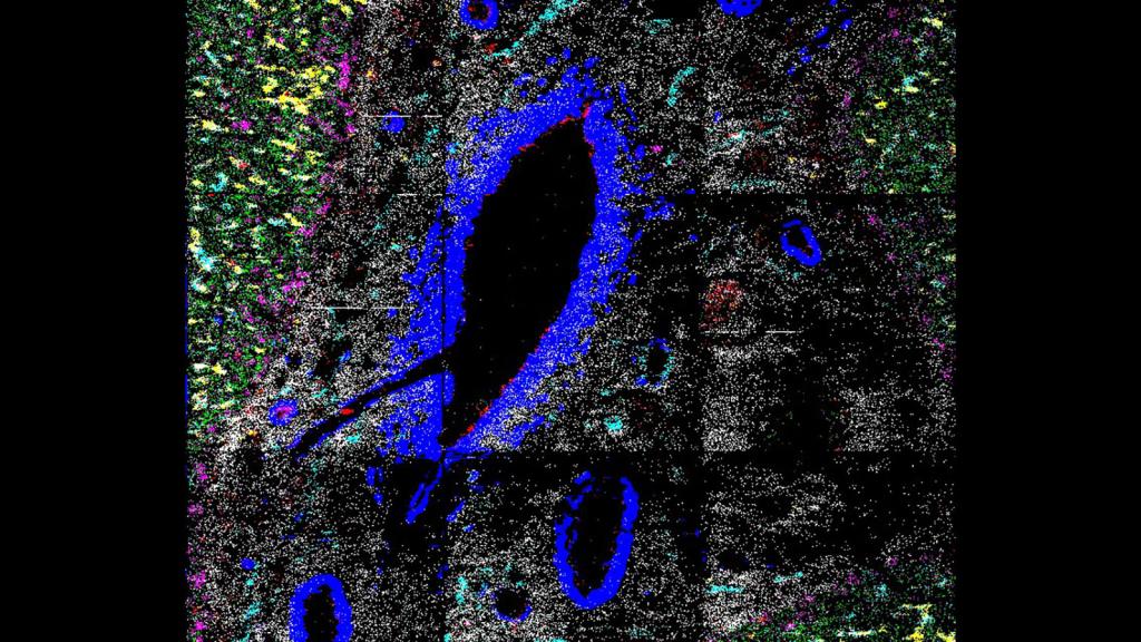
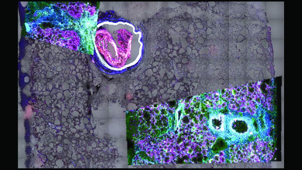
MIBI-TOF image by Dr. Albert Tsai at the Bendall Lab at Stanford University of a glomerulus and surrounding tubules within a human kidney
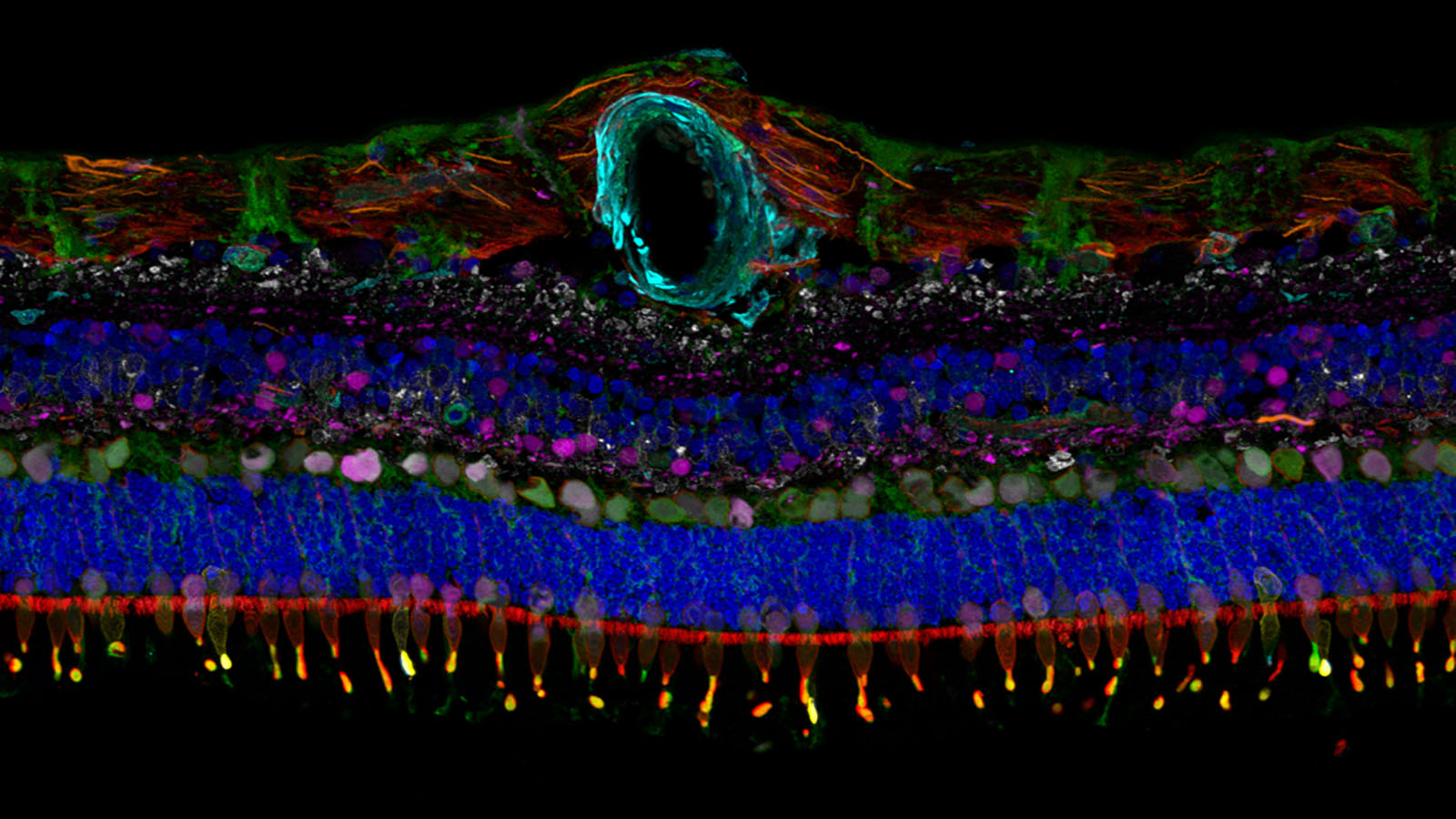
IBEX image of a human retina from Dr. Colin Chu at UCL Institute of Ophthalmology
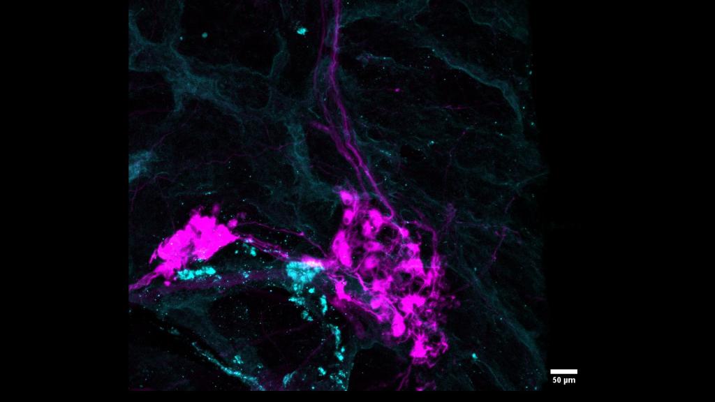
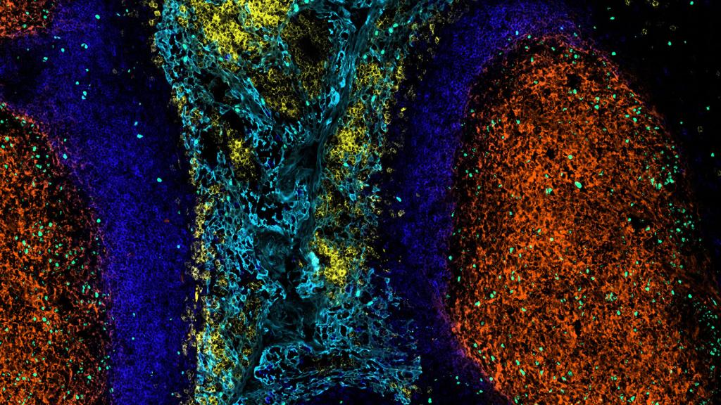
CODEX image of a neural node in the pancreas, courtsey of Dr. Angela Kruse in the Spraggins lab at Vanderbilt University
This is an IBEX image of a human spleen made by Dr. Andrea Radtke of the Germain lab at NIAID
This is a MASCima image of a human tonsil, made by Drs. Werner Muller & Andreas Bosio at Miltenyi Biotec
IMS image of a pediatric liver, courtesy of Dr. Hua Tian
CODEX image of the left atrium from Drs. Kai Tan and Kyung Ahn at Children’s Hospital of Philadelphia
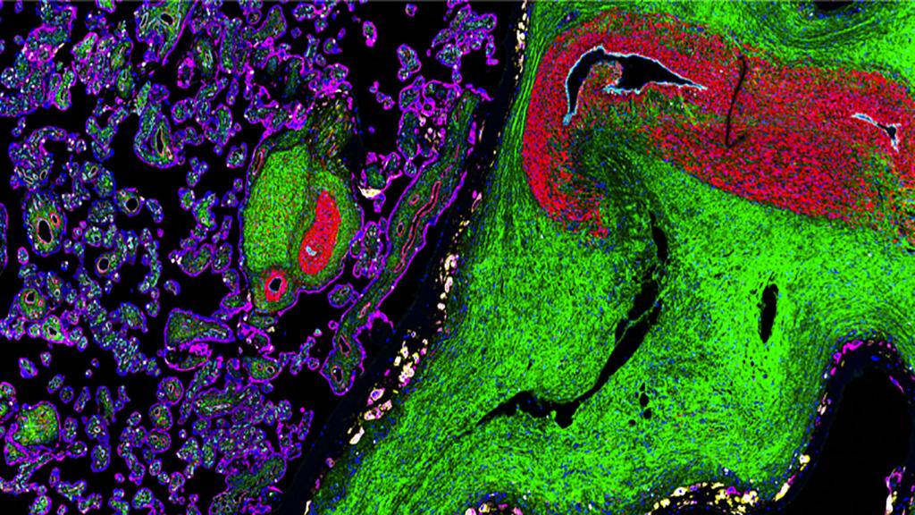
This is a MALDI-MSI & autofluorescence image of the lipids inside the central portion of the left upper lobe of the lung, showing the large bronchus and smaller bronchioles by HuBMAP researcher Dr. Brittney Gorman at Pacific Northwest National Laboratory
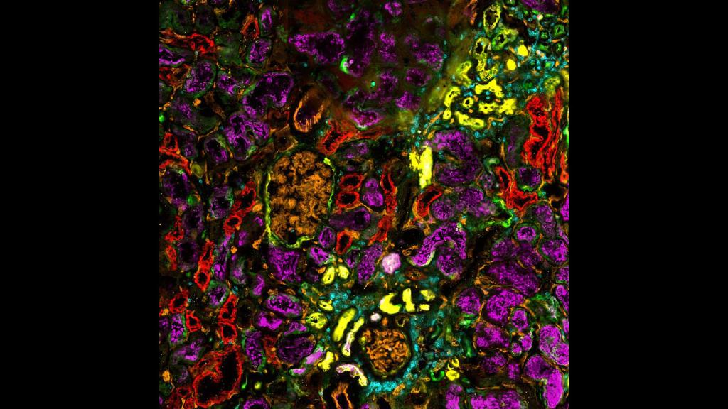
CODEX image of a human kidney from Drs. Tarek Ashkar, Angela Sabo, and Daria Barwinska at Indiana University
Organ Mapping Antibody Panel (OMAP) of the placenta from HuBMAP researchers Drs. Santhosh Sivajothi and Ramalakshmi Ramasamy at The Jackson Laboratory
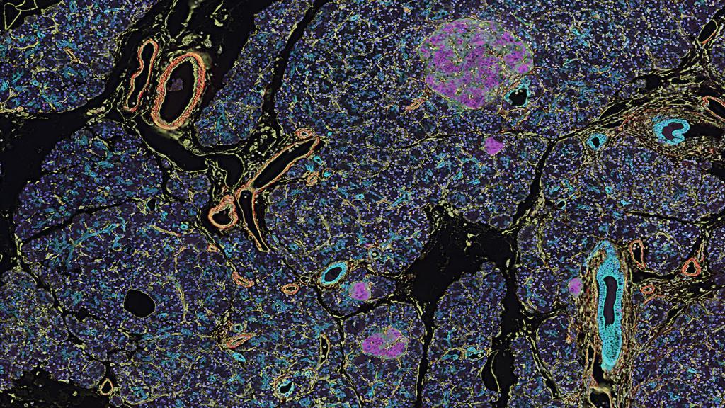
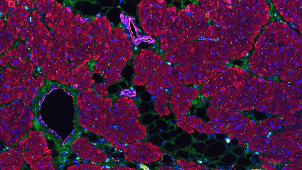
CODEX image of a healthy pancreas from Drs. Frida Björklund & Anna Martinez Casals at Emma Lundberg’s lab at Stanford
